Mykhaylo Solomonyuk
Private practice
Vilnius, 09302, Lithuania, Giedraiciu g.41-103
Cell Phone +37065385476
E- mail address. solomonyuk@gmail.com
In the article were used 2 tables and 9 illustrations.
In the 1997 graduated from Ukrainian Medical Stomatological Academy (the city of Poltava). 2000-2002 Clinical Residency at the Chair of stomatology of the National Medical Academy of Post-Graduate Education named after P.L.Shupic. 2002-2006 Full-time postgraduate courses at the National Medical Academy of Post-Graduate Education named after P.L.Shupic. 2007-2008 Completed full training course for foreign doctors on the department of orthodontics of National University of Daegu (South Korea). Membership of the American Association Orthodontists (AAO).
Summary:
In the article in presented a clinical case of treatment the patient with the frontal cant of the occlusal plane using the method of intrusion the superior lateral teeth with the anchorage on the miniscrews. Applying the last one allows to undergo a treatment without using intraoral removable and extra-oral curative orthodontic appliances, set the right position of the occlusal plane, improve the esthetic face parameters, save the molars correlation by Class І Angle. Effective control of teeth motion with the anchorage by miniscrews gives an opportunity to reduce the terms of orthodontic treatment.
Key words: miniscrews, facial asymmetry, frontal cant of the occlusal plane
Rotation of the maxillary occlusal plane is a frequent disorder of the occlusion, which is characterized by the excessive one-sided molar teeth extrusion in the maxillary bone [1]. It is common in patients with the asymmetrical mandible vertical growth, and is combined with the saggital abnormalities of the occlusion or manifest isolated[2].
Such states can lead to the functional and morphological disorders in the masticatory system. It manifest by changing the habitual mandible motions, limitation of the mouth opening, disk displacement, clicking sounds in the temporomandibular joint (TMJ). Such changes for a long time starts a process of the ankylosing the TMJ [3].
The treatment of the facial deformations with the mandibular deviation applies to the orthognatic surgery. In such clinical cases the bilateral split osteotomy is carried out. In patients with canted occlusal plane Lefort I is used [4].
Canting of the occlusal plane position, that was not complicated with the facial asymmetry, used to corrected using high-pull headgear, functional appliances, dental biteplates [5]. However, such devices are necessary no less than 16 hours a day, which is rejected by many adult patients because of the esthetical and social considerations.
Recently, for reinforcing the anchorage in case of teeth displacement, more frequent orthodontical miniscrews have been being used. It is known, that there is an efficient application in case of the anterior teeth retraction, the distal and mesial occlusion, during the control of the vertical control facial height [6–8].
The purpose of our study is to describe the clinic case of treatment the patient with the cant of the maxillary occlusal plane using miniscrews.
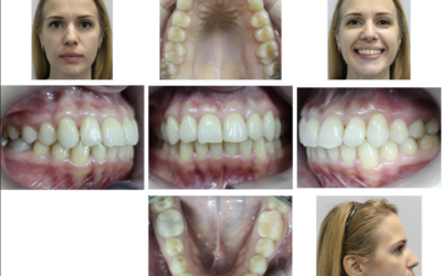
A 20-year-old patient was referred to the clinic for the orthodontist’s consultation (fig.1). During the visual examination the face was symmetrical, proportional, the mouth opening was unrestricted, any disorders on the TMJ side weren’t revealed. The profile was convex, lips were competent, the breath was nasal. The frontal facial photograph with the bite template showed the right-shifted cant of the occlusal plane.
The intraoral examination showed that the patient had a full complement of teeth. The molar relationships were class I on both sides, overjet and overbite were 1,0 and 1,0 mm, respectively. Maxillary and mandibular dental midline almost coincided with the facial midline.
The model analysis showed an absence of space on the maxillary and mandibular arches , the curve of Spee is 1 mm (fig. 2).
Panoramic radiographic showed incomplete maxillary and mandibular third molars eruption, absence of root resorption in the erupted teeth.
The lateral cephalometric analyses indicated skeletal Class I (ANB angle, 3º) with a hypodivergent growth pattern ( FH–NP 19 º , the maxillary incisor were normal inclination ( U1- SH, 108°), mandibular incisors were proclined (IMPA, 102°), (fig. 3), (table 1).
Anteroposterior (AP) cephalometric radiographs before treatment estimated used two reference lines: a horizontal line (HL), the line connecting the right and left latero-orbitale points, and a vertical line (VL), the line perpendicular to the HL through the center of crista galli (most constricted point of the projection of the perpendicular lamina of the ethmoid).
The vertical angle measurements between the HL and the bicondylar plane (Co/Co) were 0º (fig. 4). Nasal cavity floor plane (NF/NF) were rotated 5° in right-side to HL. Maxillary jugal plane (J/J) were off by 1,5º. The occlusal plane (horizontal plane passing through the molars) canted down to the right side 4°. Gonial plane were parallel to HL (table 2).
Horizontal angle measurements revealed superior midline deviation in the left side (point between the superior central incisors on the level of their incisive edges to the vertical VL plane) by 2º. Angle between the VL and mental line was off by 3º (Menton, point on the inferior boder of the symphys directly inferior to the mental protuberance). The TnS-ANS nasal septum displacement was within 1º to the VL (Anterior nasal spine (ANS), tip of the ANS below the nasal cavity and above the hard palate. Top of the nasal septum (Tns), the highest point on the superior aspect of the nasal septum).
TREATMENT OBJECTIVES
The following treatment objectives were established:
- Relieve the leveling in both arches.
- Intrusion of molars and premolars in the right maxillary
- Correction of the occlusal plane position
- Obtaine a stable occlusal relationship
- Improve the facial and dental esthetics by establishing an esthetic smile.
TREATMENT ALTERNATIVES
For solving the assigned task several alternatives have been proposed. The first suggested on the right-sided molars and premolars intrusion, applying bite-blocks with the high-pull gear. The patient had refused using removable orthodontics intra and extra-oral appliences. The second alternative was to intrude the extruded teeth and correction of the frontal occlusal plane using minisrews. The patient chose the second of treatment plan.
We placed preadjusted edgewise appliances with 0.022×0.028-in slot to his teeth in on both arches for leveling and alignment; 0,014- in and 0,016-in nickel-titanium archwires were used for leveling in the maxillary and mandibular arches, respectively. Once leveling was complete, 2 miniscrew (Dentos “AbsoAnchor” Korea, 1,3 mm in diameter and 8 mm long) were implanted of the interradicular space between the first and second premolar and the second premolar and first molar. After leveling 0.019×0.025-in stainless steel archwires were used. For intrusion of posterior teeth an elastic chain of 150 g of force was applied from the neck of the miniscrews to the around the premolars and first molar arches (fig 5).
Seven month after intrusion with elastic chain, the right molars in the maxilla were intruded about 4 mm, the canted maxillary occlusal plane was corrected.
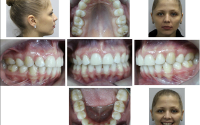
The brackets were removed after 18 months at beginning of the treatment. The posttreatment records showed that the treatment objectives were achieved. The facial photographs showed improved smile esthetics. The correction cant of occlusal plane was relieved, acceptable overbite and overjet were achieved. A class I molar relationship was maintained, and the canine relationship was improved with canine – protected occlusion. He did not report any temporo-mandibular disorder symptoms during his orthodontic treatment. Cephalometric analysis shows changes, the FMA slightly decreased from 19º to 17º. The IMPA changed from 102º to 99º. The occlusal plane has been decreased from 6º to 5º. The Z angle improved from 69º to 72º. A comparison of posteroanterior cephalometric films shows significantly improved the maxillary occlusal plane.
The posttreatment panoramic radiograph showed suitable root paralleling and no root resorption. The patient was referred to her oral surgeon for an evaluation of the extraction of his third molars (fig 6).
The posttreatment dental casts showed good intercuspation, interproximal contacts, coincident midlines (fig 7).
The posttreatment occlusion after 1 year retention under the clinic control there was no relapse, the stability of earlier received results was observed. Caine and molars positions were Class I. The absence of the dental relationship in the frontal area on the superior and inferior dentures. Dental arch have the regular shape, the saggital overjet and vertical occlusion are normal. The absence of the dysfunctional symptoms of TMJ part (fig 9).
DISCUSSION
The change of the frontal occlusal plane position can be found in patients with the facial asymmetry, as well as with balanced facial skeleton. Dental, skeletal, muscular and functional structures have the dominant impact on the occlusal plane position. But there is no exceptional influence of one definite factor, in general combined effect is observed.
The dental risk factors can be such as early deciduous teeth loss, delayed teeth eruption, congenital missing tooth or teeth, habits such as thumb sucking. Different duration of the eruption between the left and right sides of the alveolar processes can provoke to the abnormal vertical growth. If on the one side it is increased, and on the other side is reduced, mandible adapts to the lower height by shifting to this side.
The lack of the correction different vertical sizes in the growth period biases on the mandibular condyles, causing skeletal changes. The purpose of the orthodontic treatment of such patients is to eliminate different dimention in the vertical parameters between the sides by intrusion molars of non-shifted side and extrusion on the shifted.
The skeletal asymmetry also can be caused by the congenital anomalies (hemifacial microsomia or hemi-hyperplasia, but the most frequent it is a result of temporomandibular joit injury. For the symmetrical jaw growth after an injury functional appliances are used: activator Class II, bionator or others. They are made in the constructive occlusion, pulling the mandibular out to the direct contact between the superior and inferior central incisors. This new position of the jaw causes the transmission of the condyles and remodulation of the temporomandibular joint in the absence of load.
The impact of the muscle function on the maxillofacial complex formation has been the subject of active researches for many years [9]. It is proved, that the head and body position are closely connected with the functions of neck and masticatory muscles[10]. Incorrect posture, one-side chewing, habitual head tilting in one side can cause the asymmetric tension in the sternocleidomastoid and masticatory muscles. For long period of time such state leads to the asymmetric change of the skeleton, since the excessive growth starts on the one side and the less on the other. The asymmetric occlusal forms in the vertical plane between the right and left sides.
In such way, the purpose of the orthodontic treatment is to improve the discrepancy in the vertical scale by impaction molars on the unbiased side and extrusion on the biased. Here can be used the high-pull head gear and intraoral appliance, multiloop archwire mechanic with the elastic gear or miniscrews.
Correction canted of the occlusal plane with the miniscrews provides the maximum of mechanical advantages in comparison to other methods. There is no need in application extra-oral curative orthodontic appliances, the treatment begins with the arrangement of fixed systems. Simultaneous intrusion of upper molars and premolars, applying this technique, assume the minimal arch curving, decrease the time for activation the instruments[11].
Miniscrews application is the most preferable because of its simplicity in the arrangement, minimal anatomical limits during the screwing, the absence of waiting osseointegration period, direct load after setting is possible. For its stable anchorage in the osseous tissue it is important to choose the right length, diameter and set precision for the whole period of time.
The diameter of the miniscrew depends on the width of the interradicular space, which is determined on the X-ray radiograph or on the cone-beam computed tomography (CBCT) fragment. If there is enough space, it is recommended to set the miniscrew no less than 1,5 mm on the mandible and maxilla. When the space is limited with root closeness, the screw with a diameter of 1,2-1,3 mm is screwed.
The length would not have the determinative significance, because the main fixation is between the miniscrews and cortical bone. But on the maxillary it is set no less than 6 mm, on the mandibular-5 mm.
It is better to implant the miniscrew in oblique direction under the angle 30-60º to the bone surface. Such direction allows to reduce the risk of root injury during the implementation. The large volume of the cortical bone covers it, in such way it increases the adhesive force with the bone. It is better to implement the mini-screws on the gingiva area 6-8 mm from the edge. It gives a good access to the head of miniscrew for applying various variants of elastic gear.
In the available literature there are no research data about the long lasting retention of the teeth position after intrusion. But own clinical observations demonstrate the stability for a long time in the cases, where the functional occlusion is reached, right occlusal contacts, balanced function of dentofacial complex and TMJ. Indicated factors will be a good prevention of relapse.
CONCLUSIONS
- The maxillary occlusal canting can be easily treatment using miniscrew.
- The combination of miniscrew with sliding mechanics have many advantages, such as reduced treatment time, early occlusal change simplified treatment mechanics.
- For correction the maxillary canted of occlusal plane in adult it is better to use miniscrew from 1,3 mm in diameter and 8 mm long.
References
- Park YC, Lee SY, Kim DH, Jee SH. Intrusion of posterior teeth using miniscrew implants. Am J Orthod Dentofacial Orthod 2003; 123:690 – 400.
- Jeon YJ., Kim YH., Son WS., Hans MG. Correction of a canted occlusal plane with miniscrews in a patient with facial asymmetry. Am J Orthod 2006; 130:244 – 252.
- Inui M., Fushima K., Sato S. Facial asymmetry in temporomandibular joint disorder. J Oral Rehabil 1999; 26: 402 – 6.
- Kuroda S., Sakai Y., Tamamura N., Deguchi T., Takano-Yamamoto T. Treatment of severe anterior open bite with skeletal anchorage in adults: comparison with orthognatic surgery outcome. Am J Orthod 2007; 132:599 – 605.
- Bonetti GA., Giunta D. Molar intrusion with removable appliance. J Clin Orthod 1996; 30: 434 – 7.
- Kyung HM., Park HS., Bae SM., Sung JH., Kim IB. Development of orthodontic micro-implants for intraoral anchorage. J Clin Orthod 2003; 37:321 – 8.
- Соломонюк М.М. Дистализация верхних боковых зубов у взрослых пациентов с дистальной окклюзией зубных рядов с применением микроимплантатов // Ортодонтия. –2013. –№4. –С.52–58.
- Cetlin N.M., Ten Hoeve A. Non extraction treatment. J Clin Orthod 1993; 17:396 – 413.
- Solow B., Sandham A. Cranio-cervical posture: a factor in the development and function of the dentofacial structures 2002; 24:447 – 456
- Huggare J., Raustia A. Head posture and cervicovertebral and craniofacial morphology in patients with craniomandibular dysfunction. J Craniomandib Prac 1992; 10:173 – 7.
- Takano-Yamamoto T., Kuroda S. Titanium screw anchorage for correction of canted occlusal plane in patients with facial asymmetry. Am J Orthod Dentofacial Orthop 2007; 152:237 – 242.

Fig 1. Pretreatment patient’s photographs

Fig 2. On dental casts analyses


Fig 3. Lateral cephalometric: a – pretreatment; b-posttreatment


Fig 4. Posteroanterior cephalometric: a- pretreatment; b- posttreatment

Fig 5.Two miniscrews were implanted in the right side of the maxillary

Fig 6. Posttreatment patient’s photographs

Fig 7. Posttreatment dental cast.


Fig 8. Panoramic radiographs: a- pre, b-posttreatment

Fig 9. After 1 year retention patient photographs.
Table 1.Cephalometric measurements
| Measurement Norm Pretreatment Posttreatment |
| SNA 82 83 83 SNB 80 80 80 ANB 2 3 3 FMA 25 19 27 PFH/AFH 0,8 0,7 0,7 FH к Occ P 10 6 5 Ui к FH 114 108 111 IMPA 90 102 99 Z-angle 75 69 72 |
Table 2. Vertical and horizontal dimensions in the frontal teleroentgenography
| Cephalometric index (°) Before treatment After treatment |
| Bicondylar plane (HL-Co-Co) 0 0 Nasal floor plane (HL-NF’NF) 5 0 Maxillary jugal plane (HL-J’J) 1,5 1 Occlusal plane (HL-OCP) 4 0 Gonial plane (HL-Go’Go) 0 0 Superior midline (VL-isf) 2 1 Mental line (VL-Me) 3 1 Nasal septum deviation (Tns-ANS) 1 1 |


0 Comments