Mykhaylo Solomonyuk
Private practice
Vilnius, 09302, Lithuania, Giedraiciu g.41-103
Cell Phone +37065385476. E- mail address. solomonyuk@gmail.com
In the article were used 1 tables and 9 illustrations.
In the 1997 graduated from Ukrainian Medical Stomatological Academy (the city of Poltava). 2000-2002 Clinical Residency at the Chair of stomatology of the National Medical Academy of Post-Graduate Education named after P.L.Shupic. 2002-2006 Full-time postgraduate courses at the National Medical Academy of Post-Graduate Education named after P.L.Shupic. 2007-2008 Completed full training course for foreign doctors on the department of orthodontics of National University of Daegu (South Korea). Membership of the American Association Orthodontists (AAO).
Abstract:
In this article we presented clinical case of treatment patient withbimaxillary protrusion. She had Class II molars relationship and Class I canines. Merrifield Z-angle was 65°, crowded in the lower arch was -7mm, overjet was 6mm, protrusion of lower incisor IMPA was 104°. Was suggested extraction ptotocol treatment. The first premolars in the lower arch and first molars in the Upper Arch was chosen to improve crowding, lip protrusion and excessive overjet. Incisor retraction performed sliding mechanic with microscrew implants. After treatment we observed positive esthetic change. The Z-angle was improved. To achieve a satisfactory occlusion, normalizing the overjiet and overbite. The mandibular incisor were uprighted. The canine and the molars have an Angle’s Class I occlusion. Using microscrew implants to allow of upper incisor intrusion and correction gummy smile. The occlusion and the facial profile were almost stable after 1 year of retention.
Кey words: bimaxillary protrusion, microscrew implants, extraction of premolars, class I Engle.
Certainly! The term “bimaxillary protrusion” was introduced by Dr. Calvin Case in 1897. He described it as a condition where both upper and lower dental arches are positioned forward, related to other facial bones. Clinically, this manifests as protruding lips and a reduced chin profile 1. In 1926, Dr. Paul Simon proposed the term “bimaxillary protrusion” as a synonym for this condition. He further classified it into dental (where the crowns of front teeth are inclined vestibularly) and alveolar (involving anterior positioning of the bone bases) subgroups. Alveolar protrusion can be either combined with frontal dental protrusion or associated with anterior dental retrusion 2. Dr. Charles Tweed believed that most cases of bimaxillary protrusion are of dental origin rather than skeletal, contrary to earlier beliefs. He even proposed classifying them separately as Class IV according to Angle’s classification. He observed these conditions after incorrect orthodontic treatment, where central incisors were significantly vestibularly inclined. The researcher asserted that the ideal position of lower incisors relative to the mandibular plane after treatment should fall within the range of 88–90 degrees. During treatment, it’s also essential to avoid excessive expansion of dental arches, especially in the lower canine area. With such incisor positioning, even anterior alveolar protrusion appears as a flat, straight facial profile without soft tissue convexity labially [3].
The etiology of bimaxillary protrusion involves both genetic predisposition and local factors. Among these, mouth breathing, harmful tongue and lip habits, and tongue volume play a dominant role. Cephalometrically, patients with bimaxillary protrusion exhibit a shorter posterior cranial base, increased maxillary dimensions or prognathism, and a mild Class II skeletal pattern. Additionally, there is a reduction in anterior and posterior facial height, along with facial plane rotation [4].
Patients with such anomalies are treated by removing premolars. This allows reducing the convexity of soft tissues in the facial profile, correcting the gingival smile, and decreasing the undesirable vestibular inclination of upper and lower incisors [5]. However, achieving similar treatment results using the traditional straight-wire technique is quite challenging. The main problem is the lack of stable anchorage when closing extraction spaces after premolar removal [6]. As a result, unwanted mesial movement of lateral teeth, which were chosen as support, may occur. Thus, frontal tooth retraction is performed to an insufficient extent, which may not impact lip position and consequently does not improve the facial profile [7].
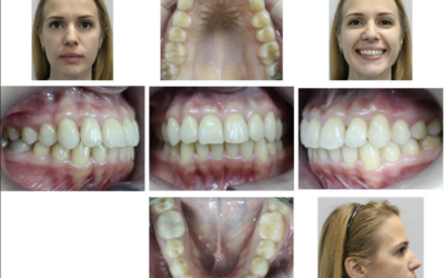
In our clinical case of treating a patient with bimaxillary protrusion, we propose a method for moving frontal teeth using microscrews implants as anchorage. The 28-year-old patient presented with complaints of unattractive tooth positioning in the upper and lower jaws and lip protrusion (Fig. 1). Upon external examination, the face appeared symmetrical, proportional, and mesiocephalic in type.

The profile is convex, lips are not closed at rest, and breathing is mixed. There is an acute nasolabial angle. The prominence of the mental crease is evident. Intraoral examination revealed that the mucous membrane of the vestibule of the oral cavity is unchanged in color, with a depth of 5 mm. The attachment of the upper lip frenulum is at a distance of 4 mm from the interdental papilla and has sufficient length without restricting lip mobility. The mucous membrane in the cheek, floor of the mouth, palate, and tongue shows no visible pathological changes. The lingual frenulum has normal attachment and does not limit tongue movement. No hypertrophy or inflammatory processes were observed on the posterior pharyngeal wall. The midlines of the upper and lower dental arches coincide. The molar relationship corresponds to Angle Class II, and the canine relationship is Angle Class I.
The analysis of plaster models revealed a deficit of space in the upper dental arch by 1 mm and in the lower dental arch by 7 mm. The Spee’s occlusal curve measures 3 mm (Fig. 2).

On the Panoramic, there were no resorptions or inflammatory changes. Absence of teeth 14 and 24 (Fig. 3).


Рис. 3. Pretreatment panoramic radiographs
The cephalometric analysis (tele-radiography) before treatment in the lateral projection revealed skeletal Class II, ANB angle of 7º (formed by the deepest point on the upper jaw – A, the junction of the nasolabial suture – Nа, and the deepest notch on the lower jaw – В), SNA angle of 84º (angle between the cranial plane SN and point A on the upper jaw), SNB angle of 77º (angle constructed by the intersection of the SN plane and point B on the lower jaw; Fig. 4). The mandibular plane angle (FMA) was 26º (formed by the mandibular plane – FH).


Fig 4. Pretreatment lateral cephalometric
The inclination of the upper incisors U1 to FH (angle formed by the axis of the upper central incisors U1 to the Frankfurt horizontal FH) is 114°. The IMPA (angle between the axis of the lower central incisors and the mandibular plane) has been increased to 105°. The Occ P to FH angle (angle between the occlusal plane and FH) corresponds to -14°. The proportional relationship between anterior occlusal height and posterior occlusal height, expressed as the PFH/AFH index, is 0.7. The Z-angle has been reduced to 65° (Table 1).
Table 1.Cephalometric measurements
| Measurements Norm Pretreatment Posttretment Retention |
| SNA 82 84 83 83 SNB 80 77 77 77 ANB 2 7 6 6 FMA 25 26 25 25 PFH/AFH 0,8 0,7 0,7 0,7 FH к Occ P 10 14 8 8 Ui to FH 114 114 107 107 IMPA 90 105 104 104 Z-angle 75 65 72 72 |


0 Comments